10+ diagram of replication fork
The proliferation of all organisms depends on the coordination of enzymatic events within large multiprotein replisomes that duplicate chromosomes. Complete diagram of a replication fork in bacterial DNA Drag the appropriate labels to their respective targets.

Draw A Labelled Diagram Of A Replicating Fork Showing The Polarity Why Does Dna Replication Occur Within Such Forks
The place where the two strands begin unwinding is called the replication fork.

. Adenine A cytosine C. Formation of Replication Forks Before DNA can be reproduced it must first be unzipped into two single strands. DNA replicates in a semi-conservative manner in which each individual strand is copied to form a new molecule of DNA.
Learn vocabulary terms and more with flashcards games and other study tools. Functions of DNA Replication DNA replication process has four major steps. 8600 Rockville Pike Bethesda MD 20894 USA.
Start studying Replication Fork Diagram. National Institutes of Health. Draw a labelled schematic diagram of a replication fork showing continuous and discontinuous.
Step- 1 Unwinding of the DNA strands and formation of replication forks Step- 2 Priming of the. Label the components first blue and then the strands pink. LIVE Course for free.
Steps of DNA Replication Step 1. Replication Fork The replication fork is a region where a cells DNA double helix has been unwound and separated to create an area where DNA polymerases and the other enzymes. The Function of the Replication Fork The replication fork is the area where the replication of DNA will actually take place.
DNA Replication Steps. Synthesis of leading and lagging strands. DNA replicates in a semi-conservative manner in which each individual strand is copied to form a new molecule of DNA.
The replication fork is moving towards the left side of the diagram. There are two strands of DNA that are exposed. The two strands can be labelled with isotopes using substrates that.
The template for the synthesis of the lagging strand. 1 mark Direction of fork movement 5 b. Continuous and discontinuous in the diagram drawn.
In the following diagram of a replication fork which DNA strand a or b is a. Replication Fork In the diagram can see one replication fork. These strands can be used to make new complementary strands called the leading strand and lagging strand.
National Library of Medicine. The top strand will be used as a template to synthesize. The two strands can be labelled with isotopes using substrates that.

K Nex Education Dna Replication And Transcription Set 525 Pieces Ages 10 Science Educational Toy History Lesson Plans Dna Lesson Plans Dna Lesson

The 10 Percent Energy Rule Studiousguy

Dna Replication Fork Definition Overview Video Lesson Transcript Study Com

Draw A Labelled Schematic Sketch Of Replication Fork Of Dna Youtube

There S More To Life Than Http Vernemq A High Performance And Distributed Mqtt Broker

Draw A Labelled Diagram Of A Replicating Fork Showing The Polarity Biology Shaalaa Com
Replication Fork Y Fork Intermediate Molecular Biology
Phosphorylation Of The Mbf Repressor Yox1p By The Dna Replication Checkpoint Keeps The G1 S Cell Cycle Transcriptional Program Active Plos One

Our Blog Posts

Draw A Labelled Diagram Of Replicating Fork

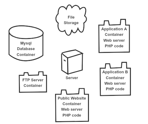
Clustered Web Applications Mysql And File Replication Roojsolutions

The Replication Fork Flashcards Quizlet

A New Class Antibacterial Almost Lessons In Drug Discovery And Development A Critical Analysis Of More Than 50 Years Of Effort Toward Atpase Inhibitors Of Dna Gyrase And Topoisomerase Iv Acs Infectious Diseases
Which Of The Two Dna Strands Is Used As A Template For Dna Replication Quora

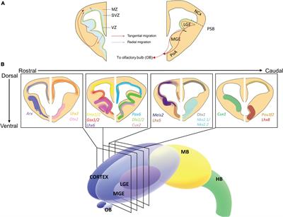
Frontiers Genetic Regulation Of Vertebrate Forebrain Development By Homeobox Genes

Eukaryotic Dna Replication Wikiwand

Tolerance Of Deregulated G1 S Transcription Depends On Critical G1 S Regulon Genes To Prevent Catastrophic Genome Instability Sciencedirect